Basic techniques for treating the avian patient

The first step to treating any injured or sick bird is proper restraint. Developing methods for restraining uncooperative birds takes time and experience. Birds possess complete cartilaginous tracheal rings, so “strangling” a bird is not a serious risk factor. However, birds breathe by moving their chest and abdomen; therefore, over-constriction of the body during restraint can cause oxygen deprivation. Birds in danger of oxygen deprivation and death while being restrained will display the following signs:
- Persistent panting and open mouth breathing
- Not attempting to bite when the head is released
- Not biting at a towel placed in its mouth
- Weak grip with both feet
If the bird shows any of these signs it is best to release and observe it.
Venipuncture
Venipuncture on psittacine birds is usually performed on the right (larger) jugular vein. For birds less than 250 grams, it is often easiest to restrain them yourself. The wing vein (basilic vein, often referred to as the brachial or ulnar vein) can also be used. Venipuncture of the wing vein tends to be uncomfortable, can create a hematoma, and in birds with calcium deficiency, may cause a wing fracture. However, many practitioners use this vein successfully.
The recurrent metatarsal vein in psittacines is quite short, and difficult to isolate in birds under 300-400 grams. Also, although hematomas are uncommon here due to the lack of metatarsal subcutaneous space, bleeding from the venipuncture site is common and a pressure wrap will be needed post-venipuncture.
Toenail trims should not be used to collect blood due to the inaccuracy of samples taken from this site. As well, it may cause discomfort to the bird, and put it at risk of septicemia if the toenail cut is high enough to allow the blood to free-flow.
Hemorrhage and hematoma formation are possible sequelae following venipuncture. In small birds this can be a major concern, and is sufficient reason to avoid taking the calculated maximum blood volume for diagnostics. Generally, maximum blood volume for withdrawal is calculated as 1% of the body weight. For example, a good-sized cockatiel can withstand a maximum of 1 ml of blood withdrawn. A 1 kg blue and gold macaw can withstand 10 ml maximum of blood removed.
Blood pressure in birds is higher than in mammals, and elevates markedly with stress. Many people apply pressure to the venipuncture site for a full 30-60 seconds after withdrawal of the needle. While this helps impede the blood seepage, the restraint also causes the blood pressure to stay elevated, increasing the likelihood of continued bleeding. Therefore, some practitioners will elect to replace the bird in its cage immediately after venipuncture is completed, if no obvious venous laceration has occurred.
Fluid administration
Subcutaneous fluids are usually given in one of three locations:
- Over the back
- Over the breast (pectoral muscles)
- In the inguinal area
When administered over the back (dorsally), the advantage is that the bird, if debilitated, can remain standing without restraint during fluid administration. Using a butterfly catheter can be helpful. The danger is that if the needle penetrates too deeply, damage to underlying organs can occur. Specifically, if done dorso-cranially, the lungs could be penetrated, while dorso-caudally the kidneys are at risk. Therefore when administering fluids in this location it is critical upon initiating injection to observe for an immediate subcutaneous bleb of fluid to form, confirming subcutaneous positioning.
Over the breast muscle is generally the safest location for the administration of subcutaneous fluids. It is still important to ensure that a bleb forms and the fluids are going subcutaneously, to prevent muscle penetration and discomfort.
Many practitioners use the inguinal route successfully. The stretching of the skin seems to cause some temporary discomfort, but it may be that this is the same when given in other locations, only more obvious when given here since the bird often limps afterwards.
Fluids IO or IV
As with all species, the availability of rapid intravenous access can be a lifesaver. However, the insertion of a port for I.V. and I.O. access will be stressful to the bird if it is mentally alert. The trade-off for obtaining intravenous access in a conscious bird is increased stress to the patient.
Crop feeding
Crop feeding is a skill that takes time and practice to learn. It can be demonstrated with images, but much like catheter placement, it requires repeated physical manipulation to successfully accomplish this in all sizes, species, and temperament of birds, without causing harm. Damage to the retropharyngeal area can occur when excessive force is used. Accidentally introducing the crop feeder into the trachea is not uncommon. Many birds (especially cockatoos) will still be able to scream with a crop feeder in the trachea. Even more commonly, backflow of the crop feeding material into the pharynx, and subsequent aspiration, may occur.
Avian emergency and triage
Immediate emergency treatment for the moribund bird
The first step is to put the bird into a warm, humidified, and oxygenated environment.
If the bird is minimally responsive, palpation of the keel and sterno-pubic area, without moving the bird, may be accomplished. Emaciation indicates chronicity, and increased sterno-pubic distance (abdominal distention) narrows the differential diagnosis. In the absence of these findings, more acute disease is likely.
Presentations other than moribund
When the bird is in the exam room with the owner, you should enter slowly, and sit down to discuss the history with the owner while also observing the bird at rest. Perform a ‘hands-off’ physical exam, which consists of observing the bird in its cage, noting respiration, mentation, grip, and posture, prior to touching the bird. Discuss the differentials, diagnostics, and treatment plan with the owner.
Seriously ill but currently stable avian patient
Birds in this category will be fluffed with a weak grip at rest. They will be temporarily able to respond to stimulation by smoothing their feathers and looking alert, but
they won’t be able to maintain this posture, and will return to being sleepy and fluffed.
Many owners don’t realize how sick their bird is, so it is important for the veterinary team to make sure they understand: proceed slowly and step-wise through the physical exam, diagnostics, and treatment.
Hospitalization
For sick birds that are still standing, especially those that have had blood loss, food and water should be readily accessible.
Perches should be removed, since a sick bird may sit perched without the energy needed to climb down to their food and water.
Seed, millet spray – whatever they will eat – should be offered. Proper diet is important, but diet conversion should be left until the bird has recovered.
The application of pressure wraps, bandaging, application of styptic, etc., will raise the bird’s blood pressure, increase bleeding, and will lead to increased stress and increased oxygen demand. For treatment, less is often more.
The bird should be placed in a quiet, warm, dark incubator, be provided with food and water, and left alone (note: anxious birds may benefit from midazolam).
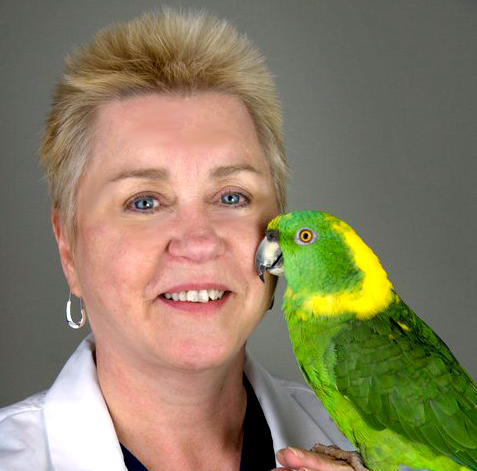
Dr. Lightfoot received her DVM from the University of Missouri in 1980, and has been practicing in Florida since 1980. She is ABVP-Avian specialty boarded and past president of ABVP. She served as a consulting veterinarian for the Suncoast Seabird Sanctuary and on the Florida Board of Veterinary Medicine, as well as the Chair from 1997-1999. She is the author and editor of two exotic medicine textbooks, ‘Clinical Avian Medicine’ and ‘Exotic Pet Behavior’. She lectures frequently on avian and exotic medicine and surgery both nationally and internationally. Dr. Lightfoot is the recipient of numerous prestigious awards for her work with exotics.
This article is based on Teresa Lightfoot’s presentation at the Western Veterinary Conference in Las Vegas, NV.CVT


